SIRT2与Bcl-2及Caspase-3在乳腺癌中的表达及其与临床病理特征的关系
梅思思1,田世维1,段彦林1,徐澍2(通讯作者)
1.贵州医科大学临床医学院病理学教研室,贵州贵阳550004; 2.贵州医科大学附属医院病理科,贵州贵阳550004
- DOI:
10.13738/j.cnki.acc.qklw60536
- 专辑:
科学Ⅰ辑;信息科技
- 专题:
信息、科学;综合科技
- 分类号:
G90;N92
[摘要]目的:通过检测SIRT2、Bcl-2、Caspase-3蛋白在乳腺癌中的表达,探讨其在乳腺癌中的表达、相关性及与乳腺癌的临床病理特征的关系。方法:采用免疫组化EnVision二步法检测384例乳腺浸润性癌和31例纤维腺病组织内SIRT2、Bcl-2、Caspase-3的蛋白表达情况。结果:乳腺癌中SIRT2、Bcl-2蛋白的高表达率高于纤维腺病,Caspase-3蛋白高表达率低于纤维腺病,SIRT2蛋白、Bcl-2蛋白在TNM分期中Ⅲ-Ⅳ期高表达率较高(P<0.05)。分子分型中SIRT2蛋白在LuminalB1型中的表达最高,Bcl-2蛋白在LuminalA型表达最高,Caspase-3蛋白LuminalA型乳腺癌中表达最低(P<0.05)。乳腺癌中SIRT2蛋白与Bcl-2蛋白表达呈正相关,与Caspase-3蛋白表达呈负相关(P<0.05)。结论:乳腺癌中可能存在SIRT2蛋白表达上调,并影响着凋亡相关蛋白Bcl-2蛋白、Caspase-3蛋白变化,从而抑制乳腺癌细胞凋亡发生,并影响乳腺癌组织学分级及TNM分期、分子分型等临床病理特征。
[关键词]乳腺肿瘤;SIRT2蛋白;Bcl-2蛋白;Caspase-3蛋白;细胞增殖;信号通路
The expression of SIRT2, Bcl-2 and Caspase-3 in breast cancer and their relationship with clinicopathological features
MEI Sisi1,TIAN Shiwei1,DUAN Yanlin1, XU Shu2
(1.Pathology Teaching and Research Section, Clinical Medicine, Guizhou Medical University, Guiyang 550004, Guizhou,China; 2.Department of Pathology, the Affiliated Hospital of Guizhou Medical University, Guiyang 550004, Guizhou,China)
[Abstract]Objective By detecting the expression of SIRT2, Bcl-2 and Caspase-3 protein in breast cancer,To explore the relationship between breast cancer and its clinicopathological features.Methods The clinicopathological features of all patients diagnosed as breast invasive ductal carcinoma in the Department of pathology, Affiliated Hospital of Guizhou Medical
from January 1, 2012 to December 31, 2017 were collected,double-blind reexamination of pathological diagnosis and histopathological grading of invasive ductal carcinoma of breast,molecular typing was classified according to Guidelines and Specifications for Diagnosi
and Treatment of Breast Cancer of China Anti-Cancer Association (2019 Edition).The protein expressions of SIRT2, Bcl-2 and Caspase-3 in 384 cases of invasive breast cancer and 31 cases of fibroadenosis were detected by immunohistochemical EnVision two-step method. Results The high expression rate of SIRT2 and Bcl-2 protein in breast cancer was higher than that in fibroadenosis, and the high expression rate of Caspase-3 protein was lower than that in fibroadenosis (P<0.05).The high expression rate of SIRT2 protein in TNM stage Ⅲ-Ⅳ was higher, and the high expression rate of SIRT2 protein in LuminalB1 was the highest in molecular typing (P<0.05).The expression rate of Bcl-2 protein in TNM stage Ⅲ-Ⅳ was higher, and the high expression rate in pathological grade I was significantly higher than that in grade II and III (P<0.05), and the highest expression rate in molecular typing was LuminalA (P<0.05).Caspase-3 protein was the lowest in LuminalA breast cancer (P<0.05).There was a positive correlation between SIRT2 protein and Bcl-2 protein expression, and a negative correlation between SirT2 protein and Caspase-3 protein expression (P<0.05).Bcl-2 protein was negatively correlated with Caspase-3 protein expression (P<0.05).Conclusion There may be an up-regulation of SIRT2 protein expression in breast cancer, which may affect the changes of apoptosis-related protein Bcl-2 in apoptosis program,The expression of Caspase-3, an executive protein of apoptosis, is down-regulated in breast cancer, thus inhibiting the occurrence of apoptosis of breast cancer cells and affecting the clinicopathological features of breast cancer such as histological grading, TNM staging, lymph node metastasis and molecular typing.
[Key words]Breast tumor;SIRT2 protein;Bcl-2 protein;Caspase-3protein;Cell multiplication;Signal channel
在中国乳腺癌将成为女性中最常见的癌症种类,其发病率排名第一,死亡率排名第五位[1]。现在的治疗方案将乳腺癌分级、HER-2、ER、PR、基因检测等分子指标引入预后判断系统,所以乳腺癌分子分型显得格外重要[2]。
Sirtuin2(SIRT2)是一种NAD+依赖性脱乙酰酶,已有研究发现SIRT2在有丝分裂、细胞的分化、细胞稳态、自噬中执行多种多样的功能。关于SIRT2是抑制还是促进乳腺癌的发展存在争议[3],有研究报道SIRT2在小鼠中充当肿瘤抑制因子,在人类样本中与正常癌旁乳腺组织相比,SIRT2在癌组织中表达较低[4]。也有研究报道SIRT2可通过促进乳腺癌小鼠模型中c-泛素化和降解而表现出抗癌作用[5]。Bcl-2和Bax分别是Bcl-2家族中重要的抑制和促进凋亡蛋白。在大多数肿瘤中,Bcl-2表达水平上升,而Bax表达水平下降。下调Bax或上调Bcl-2能抑制多种因素诱导的多种肿瘤细胞凋亡,反之则促进多种肿瘤细胞凋亡[6]。Caspase-3是一种半胱氨酸蛋白酶,在凋亡联级反应中是必经之路,其高表达水平可导致细胞凋亡,是凋亡中的核心。有研究显示,Caspase-3在肝癌、肺癌、结肠癌等多种恶性肿瘤中处于异常低表达,促使细胞增殖与凋亡平衡失调,细胞异常生长,参与了肿瘤发生[7]。
1.1临床资料
随访2012年1月1日至2017年12月31日贵州医科大学附属医院病理科诊断为乳腺浸润性导管癌的所有患者,收集患者相关临床病理特征资料(组织学分级、肿瘤直径、TNM分期、年龄、月经情况、有无淋巴结转移及有无复发)。乳腺癌患者均为女性,均为首次诊断,均未行放化疗及靶向治疗等。采用双盲法对乳腺浸润性导管癌病理诊断和病理组织学分级进行复诊,根据《中国抗癌协会乳腺癌诊治指南与规范(2021年版)》[8]对分子分型进行分类。最终确定384例患者样本纳入实验。收集31例贵州医科大学附属医院病理科诊断为纤维腺病患者,作为对照组。
1.2研究方法
1.2.1切片处理 采用免疫组织化学染色EnVision二步检测法,切片经脱蜡脱水后,将石蜡切片放在装满PH值为8.0的 EDTA 修复液盒子内高温高压修复,PBS缓冲溶液冲洗三次,每次3min。甩干PBS缓冲溶液,配好一抗,将配好的一抗稀释液滴加于切片上,放置于4℃冰箱内孵育过夜。取出4℃冰箱孵育过夜后的组织切片,放置于恒温室复温60min,PBS缓冲溶液冲洗三次,每次2min,滴加二抗,37℃水浴箱孵育60min。DAB显色,苏木素复染,并脱水透明封片观察。
1.2.2结果判读 SIRT2、Bcl-2、Caspase-3在胞浆中出现粗颗粒状和片状黄色和棕黄色着色为阳性,根据组织染色强度评分,无表达:0分;点状淡黄色:1分;粗颗粒状和片状黄色:2 分;棕黄色:3分。根据阳性细胞百分比计分:0 分(小于10%)、1 分(10%~25%)、2分(26%~50%)、3分(51%~75%)、4分(大于75%)。强度和百分比的乘积作为免疫组化结果评分(IRS),SIRT2:IRS≤3分低表达,≥4 分高表达。Bcl-2、Caspase-3 均为IRS≤8分低表达,≥9分高表达[9-11]。
1.3统计学方法
使用SPSS 22.0软件对所有数据进行统计分析,乳腺癌与对照组间SIRT2、Bcl-2、Caspase-3蛋白的表达水平比较采用卡方检验,同时,乳腺癌中不同临床病理特征组间SIRT2、Bcl-2、Caspase-3蛋白表达水平的比较也分别采用卡方检验。计算SIRT2、Bcl-2、Caspase-3蛋白指标两两之间的Spearman相关系数,以分析这些指标间的相关性,以P<0.05为差异有统计学意义。
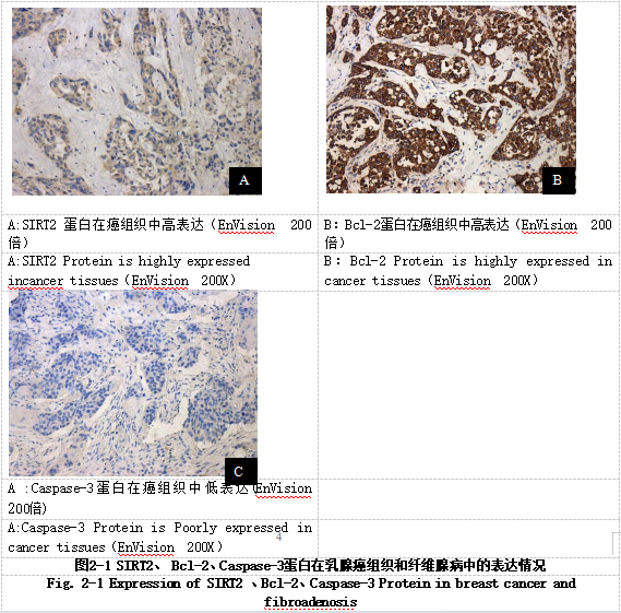
2.1 SIRT2、Bcl-2、Caspase-3蛋白在乳腺癌、纤维腺病中的表达情况(图2-1)
乳腺癌组织中SIRT2蛋白的高表达率为63.8%,高于纤维腺病的3.2%,乳腺癌中的Bcl-2蛋白的高表达率为67.6%,明显高于纤维腺病的3.2%,乳腺癌中的Caspase-3蛋白高表达率13.0%,低于纤维腺病的64.5%(P< 0.05)。
2.2 SIRT2、Bcl-2、Caspase-3蛋白表达与乳腺癌临床病理特征的关系
2.2.1 SIRT2蛋白表达与乳腺癌临床病理特征的关系 SIRT2 蛋白在 TNM 分期中Ⅲ-Ⅳ期高表达率(80.9%)较Ⅰ-Ⅱ期高表达率(58.6%)高,在分子分型中 SIRT2 蛋白在 LuminalB1 型中的高表达率(80.3%)最高,在TNBC中高表达率(35.8%)最低(P<0.05)。
2.2.2 Bcl-2蛋白与临床病理特征的关系 Bcl-2蛋白在TNM分期中Ⅲ-Ⅳ期高表达率(80.9%)较Ⅰ-Ⅱ期高表达率(66.7%)高,病理组织学分级I级中高表达率(87.0%)明显高于II级(72.4%)和III级(60.9%),在分子分型中LuminalA型表达率最高(91.9%),TNBC高表达率最低(36.8%)(P<0.05)。
2.2.3 Caspase-3蛋白与临床病理特征的关系 Caspase-3蛋白在TNM分期、组织学分级中无统计学差异(P>0.05),在分子分型中LuminalA型乳腺癌中高表达最低(7.1%),LuminalB2型中高表达率最高(17.2%)(P<0.05)。
2.3 SIRT2、Bcl-2、Caspase-3蛋白在乳腺癌中表达的相关性(见表 2-1)
本实验乳腺癌中 SIRT2与Bcl-2蛋白表达之间呈正相关(P<0.05),与Caspase-3蛋白表达负相关(P<0.05)。Bcl-2与Caspase-3蛋白负相关(P<0.05)。
表 2-1 SIRT2、Bcl-2、Caspase-3 蛋白在乳腺癌中表达的相关性
Tab.2-1 Correlation between SIRT2and expressions of Bcl-2, Caspase-3 Protein in breast cancer
Sirt2 | Bcl-2 | Caspase-3 | ||
Sirt2 | 1 | |||
Bcl-2 | 0.0469 | 1 | ||
Caspase-3 | -0.0417 | -0.0428 | 1 |
3讨论
SIRT2在细胞质中的水平在细胞周期中受到调控,它在有丝分裂过程中急剧增加,其过表达显著延长了M期[12]。有研究表明与癌旁正常组织相比,SIRT2阳性表达的肿瘤组织比例更高。本实验乳腺癌组织中SIRT2蛋白的高表达率高于对照组纤维腺病。这表明SIRT2蛋白在乳腺中的表达高于纤维腺病组织,推测SIRT2在乳腺癌的发生发展中有一定的促进作用。同时,SIRT2蛋白在TNM分期中Ⅲ-Ⅳ期高表达率较高,似乎也说明了SIRT2在肿瘤的发展过程中参与乳腺癌的演进。我们的研究结果还发现SIRT2在LuminalB1型高表达率明显增加,TNBC中SIRT2蛋白并没有表现出明显高于其他分子类型的表达上调。
Bcl-2[21]蛋白家族控制着细胞凋亡中的一个关键点,即线粒体外膜透化。Bcl-2、Bax作为其家族成员,一直备受关注。本次实验乳腺癌中的Bcl-2蛋白的高表达,在TNM分期中Ⅲ-Ⅳ期表达率较高(P<0.05),以及Bcl-2蛋白在乳腺癌不同的分子亚型中表达存在差异性,可能指导我们针对不同的乳腺癌分子分型做进一步的凋亡途径的研究。
Bax[22]信号激活Caspase-3,Capase-3表达下调足以限制上皮细胞凋亡[13],其外源性途径由细胞外配体和肿瘤坏死因子受体家族相关的跨膜死亡受体的相互作用触发,使适配器与受体结合,从而导致Caspase-8激活,活性Caspase-8进一步启动Caspase-3执行细胞凋亡[14]。内源途径是通过线粒体内膜的修饰改变线粒体跨膜电位,导致线粒体过渡孔的开放,并导致促凋亡蛋白释放到细胞质中形成凋亡小体,凋亡小体激活Caspase-9,从而导致Caspase-3激活[15]。本实验结果显示Caspase-3蛋白在乳腺癌中的表达率低于纤维腺病(P<0.05),说明在乳腺癌中Caspase-3蛋白表达下调,凋亡程序被抑制,肿瘤进一步发展。Caspase-3蛋白在分子分型中的差异性表现为LuminalA型乳腺癌中表达最低(7.1%),LuminalB2型中表达较高(17.2%)(P<0.05)。同时本次实验乳腺癌中SIRT2蛋白与Bcl-2蛋白呈正相关,与Caspase-3蛋白负相关,Bcl-2蛋白Caspase-3蛋白负相关(P<0.05)。所以我们推测在肿瘤中SIRT2、Bcl-2、Survivin蛋白起到了抑制凋亡的作用,从而促进了肿瘤的不断增殖。
结合已有的研究结果和本次研究结果,我们推测在乳腺癌组织中SIRT2影响NF-κB通路,使凋亡系列蛋白Bax蛋白与Bcl-2蛋白数量发生变化,表现为Bax蛋白表达下调,Bcl-2蛋白表达上调,进一步促进了凋亡执行蛋白Caspase-3的表达下调,导致乳腺癌细胞抗凋亡,从而促进乳腺癌发生发展。
参考文献
[1] Cao W , Chen H D , Yu Y W ,et al.Changing profiles of cancer burden worldwide and in China: a secondary analysis of the global cancer statistics 2020[J].Chinese Medical Journal, 2021, publish ahead of print.
[2] 李智勇,白雪.三阴性乳腺癌分子分型的研究进展[J].新医学, 2018, v.49(02):77-81.
[3]李翀瑶;杨宇石;孙紫君;黄瑾瑾;范梦蕾;李小虎;薄莉;徐澍.小檗碱联合阿霉素对三阴性乳腺癌MDA-MB-231/ADR细胞株增殖迁移的影响及机制[J].贵州医科大学报.2022,02:149-154
[4]Jiang C , Liu J , Guo M ,et al.The NAD-dependent deacetylase SIRT2 regulates T cell differentiation involved in tumor immune response[J].International Journal of Biological Sciences, 2020, 16(15):3075-3084.
[5]Jing H, Hu J, He B, et al. A SIRT2-selective inhibitor promotes c-Myc oncoprotein degradation and exhibits broad anticancer activity[J].Cancer Cell. (2016) 29:607.
[6]El-Mahallawy H A , El-Shakankiry N H , El-Guindy S ,et al.The Expression of Bcl2 and Bax Proteins and Their Clinical Relevance in ALL and CLL Patients[J]. 2022.
[7]王振威,肖刚,刘威,等.不同基因分型乳腺癌中Caspase-3、XIAP、Smac表达情况及与临床病理参数的关系[J].实用癌症杂志, 2019, 34(1):5.
[8] 中国抗癌协会乳腺癌专业委员会.中国抗癌协会乳腺癌诊治指南与规范(2021年版)[J]. 中国癌症杂志.2021,(10):954-1040.
[9] Taniguchi K , Karin M .NF-κB, inflammation, immunity and cancer: coming of age[J].Nature reviews. Immunology, 2018.
[10] 许安文,侯开庆,陈芳,等.乳腺癌患者Bcl-2和生存素表达与HPV感染的相关性[J].中华医院感染学杂志.2021,(2):264-267.
[11] Pratt M A C , Sau A , Cabrita M A .NF-kappaB at the crossroads of normal mammary gland biology and the pathogenesis and prevention of BRCA1-mutated breast cancer[J].Cancer Prevention Research, 2017, 11(2):canprevres.0225.2017.
[12]Chen G , Huang P , Hu C .The role of SIRT2 in cancer: A novel therapeutic target[J].International Journal of Cancer, 2020.
[13]Kuo W T , Shen L , Zuo L ,et al.Inflammation-induced Occludin Downregulation Limits Epithelial Apoptosis by Suppressing Caspase-3 Expression[J].Gastroenterology, 2019, 157(5):1323-1337.
[14]秦娟;宋国林;王建三;林虹;刘卿;陈锦云;王嫣.低强度超声辐照对人宫颈癌HeLa细胞凋亡及Casepase-12蛋白表达的影响[J].贵州医科大学学报.2019,10:1161-1166
[15]Timucin AC, Basaga H, Kutuk O. Selective targeting of antiaPoPtotic Bcl-2 Proteins in cancer[J]. Med Res Rev. 2019 ,39(1):146-175.
基金项目:组蛋白H3K56乙酰化与乳腺癌临床病理参数的关系(gzwjkj2018-1-062)
作者简介:梅思思(1987-),女,贵州安顺,本科,主治医师,研究方向:乳腺病理
相关文献推荐
- 节点文献
- 读者推荐
- 相关基金文献
- 关联作者
- 相关视频
- 批量下載